The Human Engine
Shear, Swelling, and Skin: the science of friction blisters and multi-vector mitigation in endurance sport
October 26, 2026 · 28 min read . Full open edition
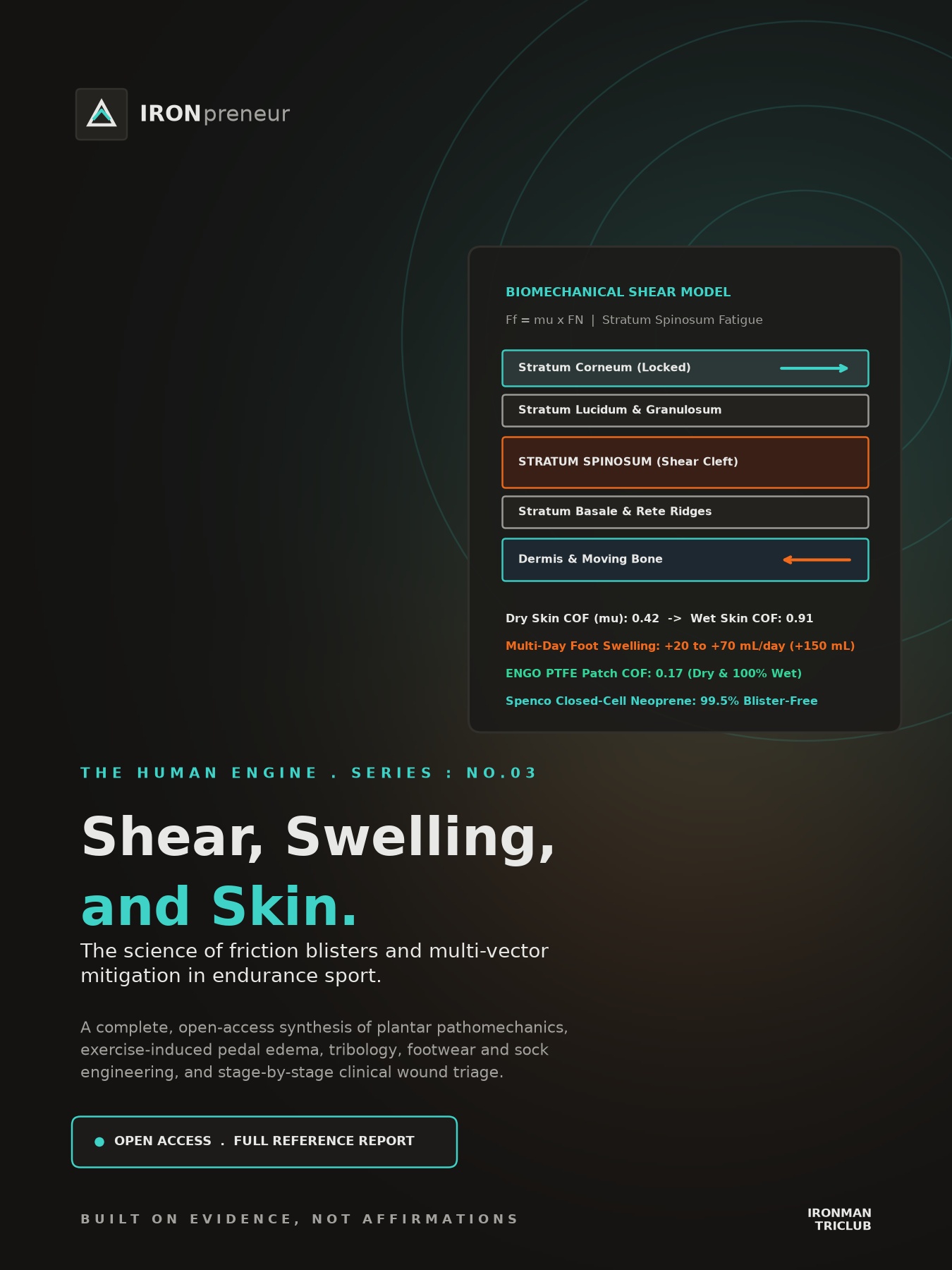
The Human Engine Series, No.03 . Complete Open-Access Reference . By Dr. Bert Grobben
During the October 2026 Singapore Perimeter Walk, stopping at 54.2 km on Day 2 was not a failure of aerobic engine, muscular endurance, or pacing. It was a mechanical failure of the plantar epidermis: severe, deep blistering on the big toes that began throbbing at rest through the night and turned every subsequent step into an open-wound infection hazard in tropical humidity. When a single dermatologic failure mode can halt an otherwise healthy endurance engine, folk wisdom is not an acceptable operating manual. This full reference edition opens the complete scientific synthesis to the public without a paywall, examining every biological, biomechanical, tribological, nutritional, and gear vector that governs blister formation and prevention. Built on evidence, not affirmations.
Medical, clinical, and field-safety disclaimer (read first)
This report is published for educational and informational purposes only. It synthesizes peer-reviewed biomedical, biomechanical, and sports podiatry literature and does not constitute medical, podiatric, dermatological, pharmacological, or clinical advice, diagnosis, or treatment. Reading or applying information from this report does not create a physician-patient or clinical relationship. Endurance events in hot, humid environments carry serious risks of tissue injury, electrolyte disturbance, and systemic bacterial infection. Any foot blister accompanied by spontaneous throbbing pain at rest, expanding redness (erythema spreading more than 10 mm beyond the blister margin), localized heat, cloudy or purulent drainage, red streaks ascending the foot or leg (acute lymphangitis), tender lymph nodes, or fever requires immediate in-person evaluation by a licensed physician. High-strength topical agents discussed in clinical literature (such as 20% aluminum chloride hexahydrate or chemical astringents) carry documented risks of irritant contact dermatitis and must never be applied to broken skin. Commercial brand names appear strictly to identify materials evaluated in published research, with zero commercial sponsorship, affiliate relationship, or endorsement by IronPreneur or Dr. Bert Grobben. See the full Disclaimers and Clinical Boundaries section at the end of this report.
The verdict, up front
Friction blisters of the feet affect 33% to 83% of participants across multi-day walks, ultramarathons, and military road marches (Wilderness & Environmental Medicine 2014; Rushton & Richie 2024, J Athl Train). For decades, athletic lore blamed a superficial triad of heat, moisture, and rubbing. Contemporary histology and biomechanics establish a fundamentally different causation paradigm: a friction blister is an intraepidermal mechanical fatigue tear within the stratum spinosum caused by repetitive horizontal soft-tissue shear deformation (Richie & Rushton 2024, J Athl Train). When sweat plasticizes the stratum corneum, the interface coefficient of friction (mu) doubles from 0.42 to 0.91 (Gerhardt et al. 2008, J R Soc Interface). Simultaneously, multi-hour endurance exercise triggers vasopressin (AVP) and aldosterone-mediated extracellular water expansion, swelling the feet by 20 to 70 mL per day and up to 150 mL cumulatively across multi-stage events (Knechtle et al. 2015, PMC4610799; Kidney International Reports 2024). Inside a fixed-volume shoe, that swelling spikes normal compressive force (FN), locks the plantar skin against the sock, and forces every gait cycle into a high-amplitude internal shear event.
Headline anchors: Richie & Rushton 2024 (PMC10783477); Rushton & Richie 2024 (PMC10783476); Gerhardt et al. 2008 (PMC2607440); Knechtle et al. 2015 (PMC4610799); Lecina et al. 2024 (PMC10831365); Lipman et al. 2016 (Stanford Pre-TAPED II).
01 . Histology, pathomechanics, and tribology
A blister is an internal shear tear in the stratum spinosum, not a surface rub
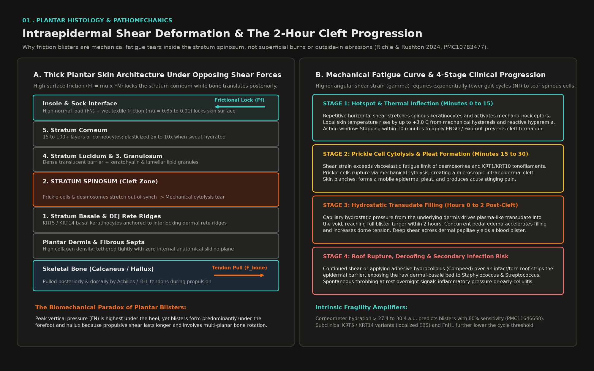
Human plantar and palmar skin possesses a specialized histological architecture adapted for high-load weightbearing and traction, with an epidermis up to 10 times thicker than skin on other anatomical regions (Richie & Rushton 2024, J Athl Train). Understanding why blisters form requires tracing the five epidermal strata from deep to superficial and observing how they are tethered to the underlying skeleton:
- 1. Stratum basale: A single layer of mitotically active cuboidal keratinocytes anchored to the basement membrane (dermo-epidermal junction, or DEJ) via hemidesmosomes, expressing basal keratin intermediate filaments KRT5 and KRT14 (Richie & Rushton 2024; Annals of Dermatology 2014).
- 2. Stratum spinosum (prickle cell layer): Multiple layers of polyhedral keratinocytes interconnected by dense cytoplasmic processes, desmosomal cadherins (desmogleins and desmocollins), internal tonofilament bundles (KRT1, KRT10, and stress-induced KRT6/KRT16), and an intercellular cement of glycoproteins and lipoproteins (Richie & Rushton 2024; J Invest Dermatol 2022).
- 3. Stratum granulosum: Flattened keratinocytes containing keratohyalin granules (profilaggrin and loricrin) and lamellar bodies that extrude waterproof barrier lipids (Richie & Rushton 2024).
- 4. Stratum lucidum: A translucent, densely packed eosinophilic layer present exclusively in thick palmar and plantar skin (Richie & Rushton 2024).
- 5. Stratum corneum: The outermost horny layer composed of 15 to 100 or more layers of anucleated, flattened corneocytes embedded in a lipid matrix, engineered to resist superficial abrasion and exfoliate gradually (Richie & Rushton 2024).
Beneath the epidermis lies the plantar dermis, which contains a substantially higher collagen content than dorsal skin and interlocks with the epidermis via deep epidermal rete ridges (Richie & Rushton 2024). Crucially, the plantar soft tissues (epidermis, dermis, superficial fat-pad septa, and deep plantar fascia) are bound together by fibrous connective tissue with zero internal anatomical sliding interface (Richie & Rushton 2024). Because the outer stratum corneum is thick and tough while the deep dermo-epidermal junction is reinforced by interlocking rete ridges, mechanical shear stress concentrates directly inside the middle layer, the stratum spinosum, where resistance to horizontal deformation is lowest (Richie & Rushton 2024).
The physics of repetitive shear deformation: Ff = mu x FN
A friction blister is neither an outside-in abrasion caused by a sock scraping skin off from the surface nor a thermal burn (Richie & Rushton 2024). Instead, blister pathomechanics are governed by the interaction between internal skeletal movement and external frictional locking, described by the classical tribological equation Ff = mu x FN, where Ff is the parallel frictional force resisting sliding at the skin-sock or sock-shoe interface, mu is the dimensionless coefficient of friction (COF) between the contacting surfaces, and FN is the normal (vertical or compressive) force pressing the surfaces together (Rushton & Richie 2024; Richie & Rushton 2024).
During human walking and running gait, plantar shear forces follow a biphasic anteroposterior trajectory (Richie & Rushton 2024). At initial contact and loading response, the foot strikes the ground at an angle, generating an anteriorly directed shear force as the calcaneus and metatarsals decelerate inside the shoe. At terminal stance and pre-swing (propulsion), elastic recoil of the Achilles tendon pulls the calcaneus superiorly while the flexor hallucis longus (FHL) and flexor digitorum longus (FDL) pull the phalanges posteriorly and plantarly (Richie & Rushton 2024).
The biomechanical paradox of plantar blisters
When mu and FN at the skin-sock-shoe interface are high, the stratum corneum remains locked in place while the underlying bone translates posteriorly or rotates. High surface friction prevents superficial slipping (rubbing) at the skin surface, which maximizes propulsive traction but forces 100% of the horizontal displacement into internal soft-tissue shear distortion (Richie & Rushton 2024). Conversely, if a low-friction interface allows early micro-sliding between the sock and shoe lining, the skin moves in synch with the bone, keeping internal epidermal shear strain below its elastic yield point. This explains why peak vertical plantar pressures (FN) are consistently higher under the heel than under the forefoot during walking and running, yet plantar blisters form far more frequently under the forefoot and toes: the propulsive shear phase at terminal stance lasts significantly longer and involves greater multi-planar bone rotation and tendon pull than the brief heel-strike event (Richie & Rushton 2024).
The mechanical fatigue curve and the 2-hour cleft timeline
Blister initiation obeys a classical materials-fatigue curve relating shear stress magnitude (tau) to the number of cycles to failure (Nf) (Richie & Rushton 2024). Higher shear distortion per step requires exponentially fewer steps to rupture the stratum spinosum, whereas lower shear distortion can be tolerated for tens of thousands of steps before reaching the fatigue limit. Histologically and clinically, the injury unfolds in three sequential phases:
- Phase 1, The Hotspot (Mechanical Nociception and Erythema): Initial repetitive shear stretches spinous keratinocytes and activates mechanosensitive and thermal nociceptors, producing a localized sensation of rubbing followed by stinging and cutaneous erythema (redness) (Richie & Rushton 2024). Local skin temperature rises by up to 3 degrees Celsius due to mechanical hysteresis and reactive hyperemia (Rushton & Richie 2024).
- Phase 2, Intraepidermal Cleavage (Prickle Cell Cytolysis): Continued shear cycles cause mechanical fatigue failure of desmosomal junctions and intracellular tonofilaments inside the stratum spinosum (Richie & Rushton 2024). Clinically, the skin blanches, forms a small mobile epidermal pleat, and registers acute stinging pain.
- Phase 3, Hydrostatic Fluid Accumulation (0 to 2 Hours): The intraepidermal tear does not fill with fluid instantaneously; rather, hydrostatic pressure from the underlying dermal capillary bed drives a thin, colorless, plasma-like transudate (with lower protein concentration than whole serum) into the void, reaching full blister turgor within 2 hours of cleft formation (Richie & Rushton 2024). If shear continues over an intact blister, the roof tears or deroofs completely, exposing the raw, nociceptor-dense dermal-basale bed (Rushton Blister Blueprint 2.0). If shear extends deeper through a rigid callus or pinches dermal papillae capillaries, red blood cells extravasate into the cleft, creating a blood blister (Rushton Big Toe Blisters).
The biphasic moisture-friction curve and thermal kinetics
While moisture and heat do not cause blisters in the absence of bone movement and shear, they act as severe tribological multipliers (Rushton & Richie 2024). The plantar foot contains the highest density of eccrine sweat glands in the human body, capable of secreting 381 to 447 g/h of sweat during exercise, which routinely overwhelms the evaporative capacity of closed footwear in hot, humid tropical climates (Rushton & Richie 2024).
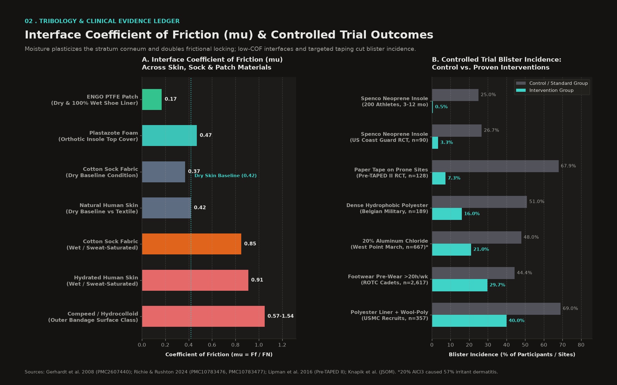
- Dry versus Moist Skin Plasticization: Water absorbed by corneocytes plasticizes and softens the stratum corneum, dropping its elastic modulus by a factor of 2 to 10, from 120 to 1,000 MPa in dry stratum corneum down to 26 to 100 MPa when wet (Gerhardt et al. 2008, J R Soc Interface).
- Real Contact Area (RCA) Expansion: As softened skin asperities flatten under vertical load, surface roughness drops twofold and the real contact area against sock fibers expands sharply, causing molecular adhesion between skin and textile to surge (Gerhardt et al. 2008).
- Doubling of the Coefficient of Friction (mu): In controlled human skin-textile friction measurements, natural dry skin exhibits a mean mu of 0.42, whereas moisture-saturated skin against wet fabric reaches a mean mu of 0.91 (averaging 0.88 plus or minus 0.08 in men and 0.95 plus or minus 0.04 in women), representing a more than twofold surge in frictional locking (Gerhardt et al. 2008). Similarly, cotton sock fabric increases from mu = 0.37 when dry to mu = 0.85 when wet (Premier Journal of Sports Science 2025).
- Prolonged Maceration Weakens Cellular Cohesion: After 4 hours of continuous moisture exposure, the stratum corneum swells threefold in thickness (and fourfold after 24 hours), degrading lateral corneocyte cohesion and accelerating mechanical fatigue failure (Gerhardt et al. 2008).
- Thermal Acceleration: Hydrated heel skin experiences a significantly faster rate of temperature rise during cyclic shear loading than dry skin (r = 0.520, p = 0.001), rapidly crossing the 3-degree-Celsius thermal inflection point that precedes epidermal cleft formation (Rushton & Richie 2024).
02 . Intrinsic susceptibility and hallux biomechanics
Why some athletes blister easily, and why the big toe is the primary failure zone
Controlled experimental blister studies since Sulzberger and Akers have shown wide inter-individual variance in the time and gait-cycle count required to blister under identical mechanical loads. Five primary intrinsic biological and biomechanical mechanisms explain why an individual athlete's skin blisters easily:
- 1. Elevated Baseline and Exercise-Induced Epidermal Hydration: In a 2024 prospective field study of 86 long-distance hikers (mean distance 455.9 plus or minus 354.5 km), 61.6% developed foot blisters (International Wound Journal 2024, PMC11646658). Corneometer measurements demonstrated that hikers who developed blisters had significantly higher plantar skin hydration (left foot 41.94 plus or minus 15.48 a.u. versus 35.77 plus or minus 10.77 a.u., p = 0.032; right foot 41.19 plus or minus 11.68 a.u. versus 36.00 plus or minus 12.85 a.u., p = 0.053), with hydration values exceeding 27.37 a.u. (left) and 30.40 a.u. (right) predicting blister occurrence with 80% sensitivity (PMC11646658). Wet socks independently correlated with blistering (p = 0.009). Athletes with naturally high plantar eccrine sweat output or primary plantar hyperhidrosis operate permanently on the steep ascending limb of the moisture-friction curve.
- 2. Keratin Intermediate Filament and Desmosomal Fragility Variants: At the molecular level, epidermal resistance to horizontal shear depends on the structural integrity of the keratin cytoskeleton in the basal and spinous layers (British Journal of Dermatology 2023; Annals of Dermatology 2014). Mild or subclinical forms of localized Epidermolysis Bullosa Simplex (EBS, formerly Weber-Cockayne syndrome) are caused by autosomal dominant missense variants in the KRT5 or KRT14 genes, and occasionally KRT6A, KRT16, plectin, or desmoplakin (Annals of Dermatology 2014; J Invest Dermatol 2022). Unlike generalized EBS, individuals with localized acral keratin variants have completely normal skin in everyday life but develop severe plantar and palmar blisters during prolonged walking, military road marches, or warm-weather endurance sport (British Journal of Dermatology 2008). Even without a clinical EBS mutation, polymorphisms in keratin filament bundling, filaggrin hydration, and dermo-epidermal junction (DEJ) rete ridge interdigitation alter an individual's shear modulus (Int Wound J 2022, PMC8762572).
- 3. Age-Related and Structural Dermal Remodeling: While baseline plantar hydration correlates inversely with age (r = -0.333, p = 0.002), aging and cumulative mechanical loading flatten the dermo-epidermal rete pegs and alter plantar fat-pad viscoelasticity, reducing the energy-dissipation capacity of the dermis during repetitive shear (PMC8762572; PMC11646658).
- 4. Dynamic Plantar Arch Collapse Under Fatigue: Three-dimensional foot scanning before and after a 10-km run demonstrates a highly significant reduction in navicular height (right foot 21.9 plus or minus 4.3 mm down to 20.3 plus or minus 3.7 mm, p < 0.001, partial eta-squared = 0.35) and navicular drop (p < 0.001), accompanied by progressive foot elongation (p = 0.007) (Frontiers in Bioengineering and Biotechnology 2024). Athletes with greater dynamic arch elongation experience larger anteroposterior shear excursion of the first metatarsal head and hallux inside the shoe with every stride.
- 5. Modality-Specific Unconditioned Epidermis: When endurance training volume is concentrated in low-shear modalities such as swimming and cycling (or short treadmill runs in air-conditioned rooms), the plantar stratum spinosum and stratum corneum do not undergo the specific shear-induced hyperplasia and desmosomal upregulation required for 8 to 10 hours per day of outdoor road walking in equatorial heat (Knapik et al., JSOM; Rushton & Richie 2024).
Biomechanics of big toe (hallux) and plantar-medial edge blisters
Severe blistering of the big toe (hallux), particularly beneath the plantar interphalangeal joint or along the plantar-medial edge, is rarely a random shoe-fit coincidence. Podiatric biomechanics identifies it as the classic clinical signature of Functional Hallux Limitus (FnHL) and compensatory toe-off mechanics (Rushton Big Toe Blisters; Quant Imaging Med Surg 2025).
| Biomechanical Driver | Pathomechanical Mechanism During Gait | Characteristic Blister Site | Primary Mitigation Vector |
|---|---|---|---|
| Functional Hallux Limitus (FnHL) | The first metatarsophalangeal (1st MTP) joint dorsiflexes normally when unloaded, but locks in the sagittal plane under closed-chain weightbearing load due to flexor hallucis longus (FHL) tenodesis or plantar fascia tension (QIMS 2025; Mentors in Orthopedics). Because the 1st MTP joint cannot hinge upward at terminal stance, propulsion shifts distally onto the underside of the distal phalanx of the hallux. | Plantar aspect and plantar-medial edge of the hallux under the interphalangeal joint and pulp (Blister Prevention). | Forefoot rocker midsole; first-ray cutout or windlass-facilitating orthotic; ENGO Two-Patch Technique under hallux and medial sidewall (Blister Prevention). |
| Abductory Twist and Foot Turnout | Restricted ankle dorsiflexion (tight gastrocnemius-soleus complex) or gluteal fatigue causes internal tibial rotation and late-stance pronation (Blister Prevention). As the heel lifts early, the foot pivots medially, forcing the athlete to roll off the medial/outer edge of the big toe rather than straight through the toe pulp. | Medial/outer plantar edge of the big toe (edge blister), frequently tearing the roof due to torsional shear (Blister Prevention). | Calf stretching and 4 to 6 mm heel-drop optimization; ENGO two-patch sidewall application; Spenco closed-cell neoprene insole (PMC10783476). |
| Hallux Valgus and Interdigital Compression | Angulation of the hallux toward the second digit or forefoot edema inside a tapered toe box compresses the lateral hallux against the medial aspect of the second toe while adjacent digits oscillate vertically out of phase (Blister Prevention). | Lateral aspect of the hallux and medial aspect of the second toe (interdigital pinch blister). | Anatomical foot-shaped wide toe box; Injinji five-toe liner socks; silicone gel toe sleeve (for prevention only, never over an existing blister). |
| Posterior-to-Anterior Toe Jamming | Downhill walking, loose midfoot lacing, or rearfoot pistoning allows the foot to slide anteriorly during loading response, impacting the distal hallux against the front toe bumper (PMC10783476). | Distal tip and subungual (under-nail) bed of the hallux or second toe. | Heel-lock (runner's loop) lacing; full thumb's width (12 to 15 mm) distal forefoot clearance; trimming toenails flush and square. |
03 . Exercise-induced foot swelling (pedal edema) and fluid dynamics
Why endurance feet swell by up to 150 mL, and how swelling triggers the shear cascade
Peripheral swelling of the feet and ankles during prolonged endurance exercise is not an incidental annoyance; it is a primary mechanical amplifier of blistering. Water plethysmography, mobile magnetic resonance imaging (MRI), and bioimpedance spectroscopy across ultra-endurance cohorts establish the exact magnitude and physiological drivers of exercise-induced pedal edema:
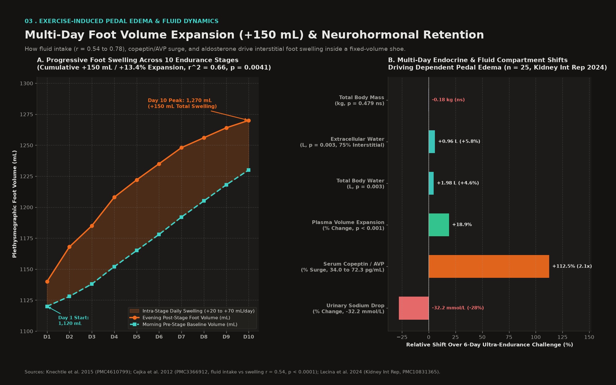
- Multi-Stage Cumulative Foot Swelling (+20 to +70 mL/day, +150 mL Total): In a 10-stage ultra-endurance triathlon study measuring foot volume via water plethysmography twice daily, morning pre-stage foot volume expanded from 1,120 mL on Day 1 to 1,230 mL on Day 10, while evening post-stage foot volume rose from 1,140 mL to 1,270 mL (r^2 = 0.66, p = 0.0041), representing a cumulative increase of 150 mL (+13.4%) (Knechtle et al. 2015, Int J Gen Med). Daily intra-stage volume increases ranged from +20 mL to +70 mL per day (r^2 = 0.52, p = 0.018).
- Direct Correlation With Fluid Intake Rate (r = 0.54 to 0.78): Across both single-stage 100-km ultramarathons (n = 76) and multi-stage events, foot volume expansion correlates strongly and positively with fluid intake rate during exercise (r = 0.54, p < 0.0001 in 100-km runners; r = 0.78, p = 0.0082 across 10-stage events) (Cejka et al. 2012, JISSN; Knechtle et al. 2015). In the 100-km cohort, foot volume change also correlated inversely with change in plasma sodium concentration (r = -0.26, p = 0.0227), proving that hypotonic fluid excess shifts water into peripheral interstitial tissues (Cejka et al. 2012). Conversely, when Ironman triathletes drank ad libitum at moderate rates (0.79 plus or minus 0.43 L/h) matched to sweat losses, serum sodium remained stable and no significant lower-limb swelling occurred (Meyer et al. 2012, JISSN).
- Weightbearing Impact versus Non-Impact Posture: Even short bouts of weightbearing treadmill walking produce measurable foot volume expansion in men (increasing from 871.0 mL to 886.2 mL, or +1.4% within a single session) due to gravitational hydrostatic pooling and impact hyperemia (McWhorter et al., J Foot Ankle Res). By contrast, 24-hour ultra-mountain bikers (non-impact, seated posture) showed zero foot volume increase (p > 0.05), confirming that upright bipedal impact loading is essential to localizing edema to the feet (JISSN 2014). Mobile MRI of runners in the Trans-Europe Footrace further documented progressive subcutaneous plantar and peritendinous edema alongside Achilles tendon diameter expansion from 6.8 mm to 7.8 mm (BMJ Open 2012, PMC3364457).
The neurohormonal cascade: aldosterone, vasopressin (AVP), and Starling forces
Why does the human body retain fluid and swell in the extremities across multi-day endurance challenges in warm climates? A landmark 2024 prospective study in Kidney International Reports tracked fluid compartments and endocrine biomarkers across a 6-day ultra-endurance challenge (Lecina et al. 2024, Kidney Int Rep):
- 1. Extracellular and Interstitial Water Expansion: Despite stable total body mass (-0.18 plus or minus 1.17 kg, p = 0.479), total body water increased by +1.98 plus or minus 1.37 L (p = 0.003), extracellular water expanded by +0.96 plus or minus 0.60 L (p = 0.003), and plasma volume expanded by +18.86% plus or minus 10.7% (p < 0.001), driving progressive ankle circumference enlargement (beta = 0.09, p < 0.001) (PMC10831365). Because 75% of sodium-retained extracellular water distributes into the interstitial space while only 25% remains intravascular, multi-day extracellular expansion directly swells dependent tissues in the feet and ankles.
- 2. Renin-Angiotensin-Aldosterone System (RAAS) Activation: Thermal stress, upright posture, and exercise-induced renal hypoperfusion stimulate aldosterone secretion, driving intense renal sodium reabsorption (PMC10831365). Urinary sodium concentration dropped by -32.18 mmol/L (p = 0.022) to reach a nadir on Day 4, while urinary osmolality surged by +250.1 mosmol/kg (p = 0.044).
- 3. Arginine Vasopressin (AVP / Copeptin) Surge: Serum copeptin (the stable C-terminal surrogate of AVP) more than doubled from 34.03 pg/mL at baseline to 72.31 pg/mL on Day 6 (p < 0.001), stimulated by both osmotic triggers (sweat-induced hypertonicity) and non-osmotic triggers (heat stress, upright posture, and interleukin-6 release from skeletal muscle stress) (PMC10831365).
- 4. Microvascular Starling Forces and Tropical Heat Vasodilation: In equatorial climates such as Singapore (30 to 34 degrees Celsius ambient temperatures), thermoregulatory cutaneous vasodilation markedly increases blood flow and capillary hydrostatic pressure (Pc) in the skin of the extremities. Combined with repetitive mechanical foot-strike inflammation and catabolic protein breakdown (evidenced by the r = 0.92, p = 0.0001 correlation between serum urea and post-stage foot volume), capillary endothelial permeability rises, driving fluid filtration into the plantar and dorsal interstitium (Knechtle et al. 2015, PMC4610799).
How pedal edema directly accelerates blistering
Pedal edema connects directly to the blister causation equation (Ff = mu x FN) through four mechanical pathways: (1) an exponential rise in normal compressive force (FN) as a 50 to 100 mL volume expansion inside a non-elastic shoe upper turns the toe box into a pressurized cylinder, spiking pressure along the plantar-medial hallux, fifth toe, and dorsal instep; (2) elimination of the sock-insole slip plane, locking the sock against the skin so internal bone movement translates 100% into stratum spinosum shear; (3) interdigital toe crowding that creates pinch shear between adjacent digits; and (4) accelerated hydrostatic cleft filling and higher blister dome tension, which increases nociceptive pain and causes earlier roof rupture.
Nutritional, pharmacological, and mechanical anti-edema protocols
- Match Fluid Intake to Thirst and Validated Sweat Rate (Never Overdrink): Because fluid intake above sweat loss is the single strongest behavioral driver of exercise-induced foot swelling (r = 0.54 to 0.78), avoid rigid schedule-based overhydration during lower-intensity walking stages where sweat rates are lower than during running (Cejka et al. 2012; Knechtle et al. 2015).
- Calibrate Sodium Concentration to Sweat Tonicity: Consume electrolyte solutions providing 500 to 700 mg/L (20 to 30 mEq/L) of sodium for standard endurance sessions, scaling to 800 to 1,200 mg/L in hot, humid conditions for heavy or salty sweaters, while avoiding uncalibrated dry salt-capsule boluses chased with excess plain water that expand total extracellular volume (ACSM Position Stand; PMC10831365).
- Strict Avoidance of NSAIDs (Ibuprofen, Naproxen, Diclofenac): Non-steroidal anti-inflammatory drugs inhibit renal prostaglandins, blunting glomerular filtration and free-water clearance while potentiating vasopressin (AVP) action. In endurance athletes, NSAIDs markedly worsen fluid retention, peripheral pedal edema, kidney injury risk, and exercise-associated hyponatremia.
- Maintain Plasma Oncotic Pressure on Multi-Day Challenges: Because prolonged catabolic exercise elevates urea and degrades plasma albumin, consuming adequate dietary protein (1.6 to 2.0 g/kg/day) and intra-event amino acids helps preserve intravascular oncotic pressure across multi-day stages (Knechtle et al. 2015).
- Intra-Walk Gravitational Unloading: During 20 to 30 minute seated breaks (every 2.5 to 3 hours), remove shoes and socks and elevate the feet to hip or heart level for 10 minutes. This immediately lowers plantar capillary hydrostatic pressure (Pc), accelerates lymphatic return, and evaporates trapped sock moisture (Gerhardt et al. 2008).
- Avoid Ankle-Cutoff Calf Sleeves During Long Walks: Wearing tight compression calf sleeves that terminate at the ankle malleoli without compressing the foot creates a distal tourniquet gradient that worsens venous and lymphatic pooling in the foot. If compression is used during or after walking, use graduated full-foot-to-calf compression socks (15 to 20 mmHg during activity or 20 to 30 mmHg during post-stage recovery), ensuring the toe box of the sock does not compress the hallux.
04 . Pre-conditioning, chemical hardening, and antiperspirants
Building shear tolerance before the start line, and the antiperspirant dermatitis trade-off
Repeated exposure to low-to-moderate horizontal shear stimulates adaptive hypertrophy of the stratum spinosum and stratum corneum, increasing desmosomal density and thickening the protective horny layer (Knapik et al., JSOM; Rushton & Richie 2024). In a study of 2,617 US Army ROTC cadets, those who wore their marching footwear for more than 20 hours per week during the two weeks prior to training had a significantly lower blister incidence than those who did not (29.70% versus 44.41%, p = 0.001) (Rushton & Richie 2024). Similarly, long-distance hikers who pre-conditioned their footwear and feet had a lower blister rate (25% versus 32%). Because cycling, swimming, and incline treadmill rucks do not replicate the flat-road propulsive shear vector of a multi-day walk, athletes must perform progressive outdoor road sessions in the exact event shoe, insole, and sock stack in comparable heat.
20% aluminum chloride hexahydrate: 45% to 56% blister reduction versus irritant dermatitis
Because plantar sweat plasticizes the stratum corneum and doubles mu, suppressing eccrine sweat output with prescription-strength topical antiperspirants has been rigorously tested in military and hiking cohorts:
- West Point Military Trial (n = 667): Cadets who applied 20% aluminum chloride hexahydrate in anhydrous ethyl alcohol to their entire feet for at least 3 consecutive evenings before a 21-km loaded road march experienced a 21% blister incidence compared to 48% in the placebo group, a 56% relative risk reduction (Knapik et al., JSOM; PMC10783476). However, 57% of the antiperspirant group developed irritant contact dermatitis (skin redness, itching, and fissuring) versus 6% in the placebo group.
- Hiker Crossover Trial (n = 96): Nightly application of 20% aluminum chloride hexahydrate for 3 days prior to hiking reduced plantar sweat output by up to 40% and cut new foot blisters by 45% versus placebo (RR 0.55, 95% CI 0.37 to 0.83), with a 12% incidence of mild irritant dermatitis (Premier Journal of Sports Science 2025).
- Why Standard Consumer Sprays and Emollient Formulas Fail: When emollients were added to 20% aluminum zirconium tetrachlorohydrex glycine to eliminate skin irritation, irritant dermatitis disappeared but 100% of the blister-prevention benefit was lost (Rushton & Richie 2024; Knapik et al. 1995, PubMed 8570998). Likewise, standard over-the-counter antiperspirant foot sprays failed to reduce plantar skin hydration or thermal rise during shear loading (PMC10783476).
- Clinical Protocol and Precaution: Only anhydrous 20% aluminum chloride hexahydrate (such as Drysol, Driclor, or Odaban) is clinically effective. Because of the 12% to 57% irritant dermatitis risk, never introduce it on race week; test tolerance 3 to 4 weeks prior by applying a thin layer at bedtime for 3 nights, washing off in the morning, and discontinuing immediately if erythema or fissuring occurs. Never apply to broken or abraded skin.
Chemical hardening (10% tannic acid), callus filing, and DEJ micronutrients
- 10% Tannic Acid Soaks: Tannic acid is a polyphenolic astringent that cross-links keratin proteins in the stratum corneum, coagulates superficial cell membranes, and reduces water uptake without causing the deep brittle fissuring of formaldehyde (PMC6855284; AJPP Tannic Acid Formulation). Daily 10-minute foot soaks in a 10% tannic acid solution (or concentrated black tea infusion) for 7 to 14 days prior to a multi-day walk firms soft, hyper-hydrated plantar skin.
- Callus Management (Avoiding the Sub-Callus Blood-Blister Trap): While thin, supple calluses protect the epidermis, thick, rigid hyperkeratotic plaques (especially along the plantar-medial pinch line of the big toe) do not deform elastically. Instead, the entire rigid callus plate shifts as a solid block, transmitting high shear stress deep into the basal layer and dermis and producing painful, deep sub-callus blood blisters that are difficult to drain. File thick calluses flat with a fine foot file and keep them supple using 10% to 20% urea cream during training, stopping urea 3 to 4 days before a multi-day event so the stratum corneum is not over-softened at the start.
- Micronutrient Support for Epidermal and DEJ Structural Proteins: For athletes whose skin blisters easily, supporting the structural proteins of the dermo-epidermal junction and spinous layer provides biological insurance (Int Wound J 2022, PMC8762572): Vitamin C (250 to 500 mg twice daily) plus collagen peptides or glycine-proline (15 g/day) to support Type IV collagen in the lamina densa and Type VII collagen anchoring fibrils; sulfur-containing amino acids (N-acetylcysteine, L-cysteine, L-methionine) and zinc (15 to 25 mg/day) for epidermal keratin disulfide cross-linking; and omega-3 EPA/DHA plus linoleic acid for stratum granulosum lamellar body ceramides.
05 . Topical products: lubricants, powders, barrier films, and tackifiers
The 90-minute lubricant rebound trap, why talc fails, and how barrier films stop skin stripping
Topical lubricants aim to convert high-friction dry or moist rubbing into low-friction boundary or fluid-film lubrication at the skin-sock interface (Rushton & Richie 2024). However, dermatological tribology reveals a critical time-dependent failure mode when lubricants are used during multi-hour endurance events:
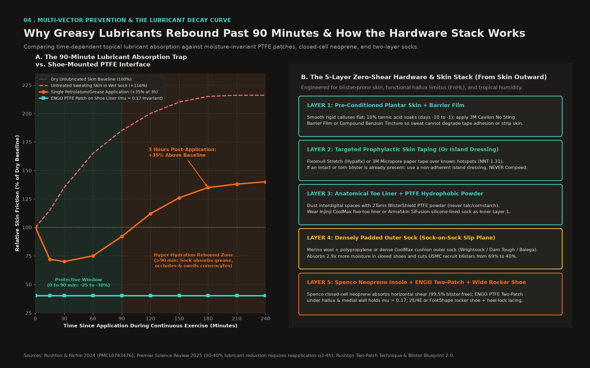
- The 90-Minute Absorption and 3-Hour Rebound (+35% Friction Surge): In controlled human skin friction studies, greasy and viscous lubricants (petrolatum, mineral oil, and glycerin) reduced mu substantially for the first 60 to 90 minutes (Rushton & Richie 2024, PMC10783476; PMC7514078). Beyond 90 minutes, the lubricant is absorbed into the sock fibers and occludes the stratum corneum, trapping eccrine sweat and hyper-hydrating corneocytes. By 3 hours post-application, skin friction rose to 35% above the unlubricated baseline (PMC10783476). Water-based or mildly greasy moisturizers increased friction almost immediately.
- Strict Reapplication Mandate: A 2025 systematic review of cutaneous friction injuries confirmed that petrolatum and silicone lubricants reduce mu by 20% to 30% in bench testing and lower field blister incidence by 30% to 40% only when reapplied every 3 to 4 hours; skipping a single scheduled reapplication doubled blister rates (Premier Journal of Sports Science 2025). High-viscosity silicone/dimethicone and wax balms (such as Trail Toes, 2Toms SportShield, Squirrel's Nut Butter, or GurneyGoo) resist washout longer than pure petrolatum but still require reapplication every 2.5 to 3.5 hours and completely preclude applying adhesive tape over the lubricated skin.
- Why Talcum and Starch Powders Fail While PTFE Powder Succeeds: British Army trials and a 5-day, 219-km ultramarathon study demonstrate that standard talcum and cornstarch foot powders provide zero reduction in blister rates or actively increase blister incidence (Rushton & Richie 2024, PMC10783476). Once sweat output exceeds the hydrophilic powder's absorption capacity, particles agglomerate into wet, abrasive clumps that elevate mu and create localized pressure points. By contrast, micronized polytetrafluoroethylene (PTFE) and polyethylene wax powder (2Toms BlisterShield Powder) is completely hydrophobic, reduces skin-surface hydration during heel shear loading, and coats sock fibers without clumping (PMC10783476).
- 3M Cavilon No Sting Barrier Film (MARSI Prevention): A fast-drying liquid acrylate terpolymer that forms a breathable, transparent, waterproof coating over the skin (3M Cavilon Technical Data). It protects fragile stratum corneum from sweat maceration, increases prophylactic tape adhesion in tropical humidity, and prevents Medical Adhesive-Related Skin Injury (MARSI): when tape is removed, the Cavilon film peels away instead of stripping epidermal cell layers (3M Surgical Tapes Guide).
- Resinous Adhesive Tackifiers (Compound Benzoin Tincture and Mastisol): Applied after an alcohol-prep wipe and allowed 30 to 60 seconds to become tacky before taping, benzoin tincture or Mastisol (liquid gum mastic) locks paper or stretch fixation tape to plantar skin across 8 to 12 hours of heavy sweating (Wilderness Medical Society; Wilderness Medicine Training Center).
06 . Taping, ENGO PTFE patches, and the hydrocolloid paradox
How prophylactic tape works, why ENGO patches belong on the shoe, and why Compeed strips blister roofs
Applying tape to the skin does not stop internal bone movement, meaning horizontal shear deformation still occurs in the soft tissues beneath the tape (Richie & Rushton 2024; SportsMAP Podcast with Rebecca Rushton). Instead, effective prophylactic skin taping works through two mechanisms: (1) presenting a smooth, low-mu outer backing so the sock glides over the tape rather than grabbing the stratum corneum, and (2) spreading horizontal shear force across a wider epidermal footprint (Rushton & Richie 2024).
- Surgical Paper Tape (3M Micropore) and the Pre-TAPED Trials: Grant Lipman and colleagues conducted two randomized controlled trials in multistage ultramarathoners. In Pre-TAPED I (2014, n = 136), where paper tape was applied to generic anatomical zones regardless of individual blister history, no protective effect was observed (Wilderness & Environmental Medicine 2014). In Pre-TAPED II (2016, n = 128), where paper tape was applied specifically to each athlete's self-reported blister-prone anatomical sites, paper tape reduced blister incidence by 40% (only 8 of 109 completers, or 7.3%, blistered under the tape versus 67.9% in untaped areas), yielding a Number Needed to Treat (NNT) of 1.31 (Stanford Medicine 2016; PMC10783476). Paper tape succeeds because it is ultra-thin, non-elastic, and has a very low outer mu, though it requires dry skin plus Cavilon or benzoin prep to survive tropical sweat.
- Non-Woven Polyester Stretch Fixation Tape (Fixomull Stretch / Hypafix / Omnifix): The endurance sports podiatry gold standard for multi-day foot taping (Blister Prevention Fixomull Guide). Fixomull conforms smoothly around complex three-dimensional contours like the big toe and heel without creasing, breathes well in humidity, resists edge rolling, and stays adhered for days.
- Rigid Zinc Oxide Tape (Leukotape P) and Synthetic Kinesiology Tape (KT Tape Pro): Ranked highest by ultrarunners in Fixing Your Feet surveys for raw adhesive tenacity (Fixing Your Feet Tape Survey). However, Leukotape P uses an aggressive zinc-oxide adhesive that must never be placed directly over an existing hotspot or fragile blister roof, as removal will strip the epidermis; KT Tape Pro offers a slick synthetic outer finish but must be applied with zero stretch at the ends to avoid epidermal tension blisters (PMC10783476).
- Moving the Slip Plane to the Shoe with ENGO PTFE Patches (mu = 0.17): Laboratory tribology testing across moisture levels from 0% to 100% by weight proved that polytetrafluoroethylene (PTFE, used in ENGO Blister Patches and ShearBan) maintains an ultra-low coefficient of friction of mu = 0.17 regardless of whether the sock is bone-dry or 100% waterlogged, compared to mu = 0.47 for Plastazote and rising friction for standard insole covers (Rushton & Richie 2024, PMC10783476; Vonhof Blister Management). For plantar-medial big toe blisters, podiatrist Rebecca Rushton's Two-Patch Technique applies one oval ENGO patch flat on the insole beneath the hallux and a second oval ENGO patch vertically up the medial sidewall of the shoe liner abutting the insole edge, allowing the sock to glide freely in both planes during propulsive roll-off without needing adhesive on the toe skin (Rushton Big Toe Blisters).
The hydrocolloid (Compeed) trap in endurance sport
Why did applying a hydrocolloid blister plaster (Compeed) during the Singapore Perimeter Walk fail to rescue the big toes? Hydrocolloids are exudative wound dressings engineered with carboxymethylcellulose particles inside an adhesive elastomer to absorb fluid from a raw, weeping, completely deroofed wound bed and form a white gel dome (Rushton Hydrocolloids for Podiatrists; Rushton Blister Blueprint 2.0). While a 40-km military march trial showed hydrocolloids could slow early hotspot progression (Premier Science 2025), placing a hydrocolloid over an intact blister or a torn-roof blister during multi-day walking in tropical heat and humidity causes three severe failures: (1) Complete roof stripping, because hydrocolloid adhesive bonds tenaciously to epidermal keratin while its outer surface has high friction (mu = 0.57 to 1.54 class), so continued walking shears the dressing and rips the fragile blister roof completely off upon shift or removal (PMC10783476); (2) Thermal edge rollback and sock melding, as tropical sweat softens unanchored edges into a sticky ridge that glues to sock fibers; and (3) Trapped maceration and infection masking when sealed over a non-sterile torn blister. The strict endurance rule: never apply a hydrocolloid over an intact or torn blister roof. Use a non-adherent island dressing (absorbent pad surrounded by Fixomull Stretch so zero glue touches the roof) for intact and torn blisters, and reserve hydrocolloids strictly for completely deroofed, raw weeping wound beds after povidone-iodine antisepsis.
| Interface / Tape Material | Outer COF (mu) | Toe Conformability | Humidity Durability | Optimal Clinical Use Case | Key Risk / Contraindication |
|---|---|---|---|---|---|
| ENGO PTFE Patch (on insole/shoe) | 0.16 to 0.17 (dry & 100% wet) (PMC10783476) | N/A (adheres to insole and shoe sidewall) | 480+ km; unaffected by sweat (Blister Prevention) | Primary shear reduction under hallux, MTP heads, and heel via Two-Patch Technique | Does not protect interdigital toe-to-toe skin contact (pair with toe socks) |
| Fixomull Stretch / Hypafix | Low to Moderate | Excellent (multi-directional non-woven stretch) (Blister Prevention) | High (breathable polyacrylate adhesive) | Prophylactic taping of toes and heels; anchoring island or hydrocolloid dressings | Slightly higher outer COF than PTFE; pair with ENGO patch for severe shear sites |
| 3M Micropore Paper Tape | Very Low (Stanford Medicine) | Moderate (non-elastic; apply in overlapping strips) | Moderate (requires Cavilon or benzoin in heavy sweat) | Prophylactic taping of athlete-specific blister-prone sites (40% reduction, NNT 1.31) | Can detach if applied to damp skin without barrier film or tackifier |
| Leukotape P (Rigid Rayon) | Moderate | Low (rigid backing creases on small toes) | Extreme (aggressive zinc-oxide adhesive) (Fixing Your Feet) | Broad flat surfaces (plantar arch and rearfoot) in wet conditions | High MARSI risk: strips fragile skin or blister roofs if applied without an island pad |
| KT Tape Pro (Synthetic) | Low | High (unidirectional elastic stretch) | Moderate to High (H2O acrylic adhesive) | Heel and arch pre-taping when applied with zero stretch at anchor ends | Applying under tension creates epidermal shear blisters at the tape ends |
| Hydrocolloid (Compeed) | High (0.57 to 1.54 class) (PMC10783476) | Moderate (thick beveled elastomer wafer) | Poor under active shear and tropical heat (melts and rolls) | Completely deroofed weeping blisters ONLY (post-event or framed with Fixomull) (Rushton Blueprint) | Never apply to intact or torn-roof blisters during walking; strips roof off on removal |
Pressure-deflective felt aperture (donut) pads and silicone gel toe sleeves
Cutting a horseshoe or circular aperture (donut) out of 5-mm to 7-mm compressed orthopedic wool felt (or stacked moleskin) and adhering it around, never directly on, a blister or hotspot shifts vertical normal force (FN) onto the surrounding perimeter (Rushton & Richie 2024, PMC10783476). Biomechanical plantar pressure studies confirm that 5-mm felt padding reduces localized peak plantar pressure by 24% to 31%, 7-mm felt donut pads reduce pressure by more than 25%, and 20-mm felt reduces peak pressure by 49%. Soft medical-grade silicone gel toe sleeves absorb internal shear deformation like an external fat pad and are effective for preventing interdigital pinch and distal hallux blisters provided the toe box has sufficient volume, but gel sleeves must never be worn over an existing or open blister during long walks because they completely occlude sweat evaporation and macerate the damaged roof (Rushton Big Toe Blisters).
07 . Sock architecture, fiber physics, and multi-layer interfaces
Why cotton fails, how Merino-polypropylene blends beat pure synthetics in closed shoes, and the two-sock slip plane
Socks govern all three mechanical variables of blistering: moisture transport away from the stratum corneum (controlling mu), vertical shock and pressure attenuation (controlling FN), and internal sock-on-sock shear absorption (Rushton & Richie 2024, PMC10783476).
- Why Cotton Is Contraindicated: Cotton is hydrophilic, absorbs 3 times as much moisture as acrylic, takes 10 times longer to dry than synthetic fibers, has low thermal conductivity (0.07 W/m.K), loses structural loft when wet, and doubles its mu from 0.37 (dry) to 0.85 (wet) (PMC10783476; Premier Science 2025). In Herring and Richie's classic double-blind RCT of long-distance runners, padded cotton socks produced twice as many blisters and blisters 3 times larger than identically constructed padded acrylic socks (PMC10783476).
- Hydrophobic Synthetics (CoolMax Polyester, Polypropylene, Acrylic, Nylon): CoolMax polyester uses a 4-channel scalloped oval cross-section that increases fiber surface area by 20% and dries 15% faster than acrylic, with twice the thermal conductivity of cotton (0.14 W/m.K), while nylon (polyamide) provides high abrasion resistance and 0.25 W/m.K thermal conductivity (PMC10783476). In a trial of 189 Belgian military recruits, a single densely padded hydrophobic polyester sock (88% polyester, 11% polyamide, 1% elastane) reduced blister incidence to 16% compared to 51% in standard wool socks and 32.3% in a cotton-wool double-sock system.
- The Closed-Shoe Evaporation Bottleneck (50% Merino Wool / 33% Polypropylene Blends): Because closed footwear in humid tropical climates restricts evaporative cooling, wicking alone cannot remove 400 g/h of sweat (PMC10783476). In a 4-day military march study (n = 37), a 50% Merino wool / 33% polypropylene blend absorbed 2.9 times more moisture into its fiber core than a 99% polypropylene sock, keeping skin significantly drier at the dorsal metatarsals and posterior heel.
- Dense Cushioning and Knit Stitch Density: When sole padding was removed in Herring and Richie's follow-up trial, thin acrylic socks showed no blister advantage over thin cotton socks (PMC10783476). Increasing knit stitch density reduces plantar shear peaks by nearly 20%, and only thick wool or acrylic cushion-sole socks maintain shock attenuation across 72 hours of loading.
- Double-Sock Layering Systems (69% down to 40% Incidence): Wearing a thin, hydrophobic polyester or nylon inner liner sock inside a dense wool-polypropylene outer sock introduces a dedicated sock-to-sock sliding interface. In a 12-week trial of 357 US Marine Corps recruits, pairing a thin polyester liner with a thick 50% wool / 50% polypropylene outer sock reduced overall blister incidence from 69% to 40% and cut severe blisters requiring medical attention from 24% down to 9% to 11% (PMC10783476; Knapik et al., JSOM). Integrated double-layer socks (such as Wrightsock Coolmesh II or Escape) achieve the same internal slip plane without bunching.
- Anatomical Five-Toe Liner Socks (Injinji CoolMax / NuWool) and Silicone-Polymer Liners (ArmaSkin): Wrapping each digit in an individual wicking sleeve eliminates skin-on-skin interdigital friction and absorbs hallux-to-second-toe pinch shear. Wearing an ultra-thin Injinji CoolMax Liner underneath a cushioned Merino-blend or synthetic outer sock combines toe separation with a two-sock plantar slip plane. Alternatively, ArmaSkin liner socks use a proprietary SiFusion hydrophobic silicone polymer inner coating that clings gently to the skin while presenting a slick nylon outer surface against the outer sock, ensuring 100% of sliding occurs between the two socks (ArmaSkin Technical Overview).
- Intra-Event Sock Rotation Hygiene: On walks exceeding 4 hours in tropical heat, swap into a fresh, dry pair of socks every 10 to 15 km (every 2.5 to 3 hours), clip damp socks to the pack exterior to dry, and rinse socks thoroughly each night so dried sweat salt crystals do not act as micro-abrasives the next morning.
08 . Footwear engineering, shear-absorbing insoles, dynamic lacing, and gaiters
Why vertical shock insoles fail (2,754 Marines) while closed-cell neoprene succeeds (99.5% blister-free)
Clinical trials reveal a decisive biomechanical distinction between insoles that cushion vertical impact (FN) and insoles that deform horizontally to absorb plantar shear (Rushton & Richie 2024, PMC10783476):
- Why Vertical Shock Absorption Alone Fails: In a massive trial of 2,754 Royal Marine recruits, replacing standard flat 3-mm insoles with 3-mm shock-absorbing insoles produced zero reduction in blisters (18.6% versus 17.2%) (Rushton & Richie 2024, PMC10783476).
- Closed-Cell Neoprene (Spenco) Shear-Absorbing Insoles: Spenco insoles are constructed from nitrogen-injected closed-cell neoprene rubber topped with a 4-way stretch nylon fabric. As the foot's bones translate during push-off, the nitrogen bubbles in the neoprene deform horizontally in synch with the skin, absorbing shear strain inside the insole rather than inside the stratum spinosum (PMC10783476). In a study of 200 athletes using a closed-cell neoprene insole in one shoe and their standard insole in the contralateral shoe for 3 to 12 months, 99.5% of feet with the neoprene insole remained completely blister-free versus 75% of control feet (PMC10783476). In an 8-week US Coast Guard recruit RCT (n = 90), only 1 recruit (3.3%) in the Spenco neoprene insole group developed blisters or calluses, compared to 4 (13.3%) in the Poron polyurethane group and 8 (26.7%) in the unpadded control group (PMC10783476).
- Anchoring Orthotics: If custom or prefabricated orthotics sit even 2 mm forward of the heel cup, their posterior edge creates severe heel-edge blisters; always anchor the underside of an orthotic to the midsole with double-sided tape (Blister Prevention Orthotics Guide).
- Anatomical Foot-Shaped Toe Boxes, Width Sizing, and Forefoot Rocker Geometry: Standard athletic shoes taper symmetrically toward the center of the forefoot, forcing the hallux laterally and pinching its plantar-medial border against the insole edge when the foot swells by 50 to 100 mL (Rushton Big Toe Blisters). Select shoes built on an anatomical foot-shaped last where the medial border remains straight (such as Topo Athletic Ultraventure, Phantom, or Atmos with a 5-mm drop and wide anatomical toe box, or Altra Olympus / Torin with a FootShape toe box), or select 2E (Wide) and 4E (Extra-Wide) lasts in cushioned road cruisers with a pronounced forefoot rocker (such as New Balance Fresh Foam X 1080 or More in 2E/4E, Brooks Ghost Max or Glycerin in 2E/4E, or Hoka Bondi/Clifton in Wide/Extra-Wide after verifying the midsole bucket wall does not rub the medial arch). Caution on zero-drop transitions: shifting abruptly from an 8 to 10 mm drop to 0 mm increases Achilles and gastrocnemius tension, which exacerbates functional hallux limitus and early heel lift; a 4 to 6 mm drop with a stiffened forefoot rocker unloads both the calf and the 1st MTP joint (QIMS 2025).
- Multi-Day Half-Size-Up Shoe Strategy: For multi-day walks exceeding 30 km/day in tropical heat, start Day 1 in a shoe with a full thumb's width (12 to 15 mm) of distal clearance and keep a half-size larger or 2E-to-4E wider backup pair ready for Day 2 afternoon or Day 3 to accommodate the cumulative +70 to +150 mL foot volume expansion (Knechtle et al. 2015).
- Four Precision Lacing Architectures and Breathable Trail Gaiters: (1) Heel-Lock Lacing (Runner's Loop) through the top auxiliary eyelets anchors the calcaneus into the heel cup to stop forward sliding and toe jamming; (2) Window (Box) Lacing skips the diagonal crossover over the dorsal instep to relieve pressure over a swelling navicular; (3) Diagonal Hallux-Lift Lacing threads from the medial front eyelet diagonally to the lateral top eyelet to lift the toe-box roof off the big toe; and (4) a Dual-Zone Surgeon's Knot halfway up the eyelets keeps the forefoot loose for toe splay while locking the midfoot, re-adjusted 45 minutes into each walk once initial hyperemic expansion stabilizes (Fixing Your Feet Lacing Guide). Top with ultralight breathable Lycra spandex gaiters (such as Dirty Girl or Altra Trail Gaiters) to block coastal sand and grit without trapping heat.
09 . Clinical wound triage, infection red flags, and return-to-swim protocol
Stage-by-stage blister management, decoding overnight throbbing, and protecting the healing epidermis
When a hotspot or blister develops during an event, clinical management must match the exact structural integrity of the epidermal roof (Rushton Blister Blueprint 2.0; Wilderness Medical Society Guidelines 2014):
| Epidermal Stage | Clinical Presentation | Immediate Cleansing & Action | Primary Dressing & Shear Offloading | Strict Contraindications |
|---|---|---|---|---|
| 1. Hotspot (Pre-Cleft) | Localized warmth (+3 C), erythema, stinging or rubbing sensation; no fluid pleat yet (PMC10783477). | Stop within 10 minutes. Remove shoe and sock, air-dry foot for 5 minutes, wipe skin with alcohol prep pad. | Apply ENGO PTFE patch to the corresponding spot on the insole/shoe sidewall AND apply Fixomull Stretch or 3M Micropore over Cavilon/benzoin to the skin (Blister Prevention). Put on dry socks. | Never wait until the next meal stop; the stratum spinosum cleft tears within 15 to 30 minutes of hotspot onset. |
| 2. Small Intact Blister (< 5 mm) | Small fluid-filled epidermal bubble; not tense or painful enough to alter gait. | Do not lance or unroof; the intact epidermal roof is a sterile biological barrier against bacterial infection (WMS 2014). | Cover roof with a non-adherent island dressing (such as Cutiplast or Primapore) so zero adhesive touches the roof (Rushton Blueprint). Offload with a 5 to 7 mm felt donut pad and apply an ENGO patch to the shoe. | Never stick tape or a Compeed hydrocolloid wafer directly onto the intact blister roof. |
| 3. Large or Weightbearing Intact Blister (>= 5 mm) | Tense fluid dome under the hallux, forefoot, or heel that will spontaneously burst under body weight. | Disinfect skin with alcohol or liquid povidone-iodine (Betadine). Lance at 2 to 3 dependent perimeter points using a sterile hypodermic needle or #11 scalpel blade (WMS 2014). Gently express fluid with sterile gauze while preserving 100% of the blister roof. | Swab liquid Betadine onto puncture sites, cover with a non-adherent island dressing framed with Fixomull Stretch, offload with a felt donut aperture pad, and apply an ENGO patch to the insole (Rushton Blueprint). | Never use a flame-heated sewing needle (deposits carbon particles); never unroof a drained intact blister; diabetic or neuropathic individuals must not self-lance. |
| 4. Torn-Roof Blister | Blister roof has split or partially peeled back, exposing part of the raw basal bed. | Irrigate thoroughly with sterile saline or clean potable water to flush grit; apply liquid povidone-iodine so it wicks under the torn flap (Rushton Blueprint). Keep clean residual roof flat over the base unless necrotic or purulent. | Cover with a non-adherent silicone mesh (Mepitel) or island dressing secured with Fixomull Stretch; offload with felt aperture padding. | Never apply a hydrocolloid (Compeed) over a torn roof; it will rip the remaining flap off and trap bacteria (Blister Prevention). |
| 5. Completely Deroofed Blister | Epidermal roof is completely gone, leaving a raw, weeping, painful dermal-basale bed (Rushton Deroofed Blisters). | Irrigate with sterile saline, disinfect with liquid povidone-iodine, and dry the surrounding intact skin. | Once walking has stopped or shear is eliminated and no infection signs exist, apply a hydrocolloid dressing (Compeed) framed around all edges with Fixomull Stretch, or a Spenco 2nd Skin moist hydrogel pad under an island dressing (Rushton Blueprint). | Never apply an occlusive hydrocolloid if there is any sign of infection (throbbing at rest, spreading redness, heat, or pus). |
Decoding overnight throbbing, infection red flags, and return-to-swim protocol
Once a blister roof tears or deroofs inside a warm, sweat-saturated shoe, the stratum corneum barrier is breached, giving skin commensals and environmental bacteria (primarily Staphylococcus aureus and beta-hemolytic Streptococcus pyogenes, or Pseudomonas aeruginosa, Aeromonas, and Vibrio species after puddle or coastal water immersion) direct entry to the dermal capillary and superficial lymphatic plexuses (Knapik et al., JSOM; Rushton Blister Blueprint 2.0).
- Mechanical Nociception versus Overnight Throbbing at Rest: Uncomplicated mechanical blister pain is load-dependent; it hurts when compressed or sheared and subsides rapidly when shoes are removed and the feet are elevated. Spontaneous throbbing through the night and pain without touch at rest indicate elevated interstitial tissue pressure inside a closed anatomical space driven by acute inflammatory vasodilation, neutrophil infiltration, or early secondary bacterial cellulitis and lymphangitis (Rushton Blister Blueprint 2.0).
- Four Clinical Red Flags Requiring Immediate Physician Evaluation: (1) Spontaneous throbbing pain at rest that persists or worsens overnight; (2) Cloudy, opaque, yellow, or purulent exudate accumulating beneath the blister roof or dressing; (3) Expanding perilesional erythema (redness spreading more than 10 mm beyond the blister margin; outline the border with a pen to track progression), localized heat, and indurated swelling; and (4) Red streaks ascending the dorsum of the foot or leg (acute lymphangitis), tender inguinal or popliteal lymph nodes, or systemic symptoms (fever, chills, malaise).
- Medical Management of Suspected Infection: If secondary bacterial infection is suspected, any occlusive hydrocolloid wafer must be soaked off and removed immediately, because sealing bacteria under an occlusive gel dome accelerates deep tissue spread. A physician will evaluate the wound for debridement of necrotic roof flaps, bacterial swabbing, targeted oral antibiotic therapy, and daily breathable antimicrobial island dressings.
- Return-to-Swim and Triathlon Training Protocol: During the active inflammatory or open weeping phase (typically Days 1 to 4 post-injury), withhold all pool and open-water swimming; chlorinated pools (which can harbor Pseudomonas and Mycobacterium marinum) and tropical open water (Vibrio and Aeromonas) macerate migrating basal keratinocytes and introduce waterborne pathogens. Once resting pain and erythema have completely resolved and a dry pink neodermis covers the base (typically Days 4 to 7+), pool swimming may resume only with a double-layer waterproof seal: place a sterile non-adherent pad over the site, seal with a wide polyurethane film dressing (3M Tegaderm or Opsite Flexifix) bordered with 3M Cavilon barrier film, avoid hard wall push-offs on flip turns, and remove the film immediately post-swim to rinse, disinfect with liquid Betadine, and air-dry.
10 . Integrated 4-phase implementation checklist
The complete multi-vector protocol from four weeks out to overnight recovery
| Phase & Window | Target Vector | Concrete Protocol & Specification |
|---|---|---|
| Phase 1: 4 Weeks to 3 Days Pre-Event | Footwear, Insoles & ENGO Setup | Select an anatomical wide-toe-box road or trail cruiser (Topo Athletic, Altra, or 2E/4E wide last) with a forefoot rocker and 12 to 15 mm distal clearance. Replace factory insoles with Spenco closed-cell neoprene insoles (99.5% blister-free in trials) and apply ENGO PTFE patches (mu = 0.17) using the Two-Patch Technique under the hallux and up the medial sidewall (PMC10783476; Blister Prevention). Wear the exact stack on road walks > 20 h/week. |
| Phase 1: 4 Weeks to 3 Days Pre-Event | Callus Filing, Tannic Acid & Sweat Control | File rigid plantar-medial hallux calluses flat and apply 10% to 20% urea cream nightly until supple (stop urea 4 days pre-event). Perform daily 10-minute 10% tannic acid (or strong black tea) foot soaks for 10 days pre-event. If plantar sweating is severe, patch-test anhydrous 20% aluminum chloride hexahydrate 3 weeks out; if tolerated without dermatitis, apply nightly for 3 evenings before the event. |
| Phase 2: Event Morning (Zero-Shear Stack) | Barrier Film, Prophylactic Tape & 2-Layer Socks | Wipe feet with alcohol prep pad, apply 3M Cavilon No Sting Barrier Film (or Compound Benzoin Tincture) to all blister-prone zones, let dry 60 seconds, and apply smooth Fixomull Stretch or 3M Micropore strips with rounded corners (3M Cavilon). Dust toes with 2Toms BlisterShield PTFE powder (never talc). Put on an Injinji CoolMax five-toe liner (or ArmaSkin SiFusion liner) as Layer 1 and a densely padded Merino-polypropylene or CoolMax outer sock (Wrightsock, Darn Tough, or Balega) as Layer 2. |
| Phase 2: Event Morning (Zero-Shear Stack) | Precision Lacing & Grit Protection | Tie shoes with a mid-eyelet surgeon's knot (loose forefoot for width expansion, snug midfoot) and a Heel-Lock (Runner's Loop) at the ankle collar. Fit breathable Lycra spandex gaiters over the shoe collar. |
| Phase 3: Intra-Event (Every 2.5 to 3 Hours) | Anti-Edema Unloading, Hydration & Sock Swap | Drink to thirst and validated sweat rate using 500 to 1,000 mg/L sodium solutions; never overdrink hypotonic fluids and take zero NSAIDs (Cejka et al. 2012). Every 2.5 to 3 hours (10 to 15 km), remove shoes and socks for 10 minutes, elevate feet to hip level to drain interstitial hydrostatic fluid, swap into dry socks, and re-tension laces for the +20 to +70 mL daily foot swelling. Stop within 10 minutes of any hotspot. |
| Phase 4: Overnight Multi-Stage Recovery | Antiseptic Hygiene, Inspection & Air Exposure | Wash feet with mild antiseptic soap, leave intact well-adhered Fixomull in place if the underlying skin is completely pain-free (or soak off gently with warm water/oil if inspecting a blister), apply non-adherent island dressings to any intact or torn blisters (never Compeed), elevate legs for 15 minutes before sleep, and wear open-toe recovery slides whenever off the road. |
Disclaimers, clinical boundaries, and disclosures
Important medical, podiatric, pharmacological, and non-endorsement disclaimers
Because this report is published as an open, un-gated public reference on the IronPreneur website and discusses clinical wound management, prescription-strength topical agents, electrolyte physiology, and medical devices, readers must review and observe the following five boundaries:
- 1. General Educational and Informational Purposes Only (Not Medical Advice): All content, biomechanical models, physiological data, and implementation checklists in this report are provided strictly for general educational and informational purposes. Nothing in this report constitutes medical, podiatric, dermatological, nutritional, or nursing advice, clinical diagnosis, or medical treatment, nor does it substitute for an in-person consultation with a qualified sports medicine physician, podiatrist, dermatologist, or registered sports dietitian. Use of this information is entirely at the reader's own risk.
- 2. Acute Bacterial Infection, Cellulitis, and Sepsis Warning: Open, torn, or deroofed foot blisters exposed to sweat-saturated socks, road dust, rainwater puddles, or natural water bodies carry a significant risk of acute bacterial skin and soft-tissue infection (including Staphylococcus aureus, MRSA, Streptococcus pyogenes, Pseudomonas aeruginosa, Aeromonas hydrophila, and Vibrio vulnificus). Spontaneous throbbing pain at rest, expanding redness, swelling, warmth, pus, red streaks ascending the limb (lymphangitis), tender lymph nodes, or fever are medical urgencies that can progress rapidly to deep cellulitis or systemic sepsis. Do not attempt field self-treatment or apply occlusive dressings over suspected infection; seek immediate medical care.
- 3. Topical Chemicals, Antiperspirants, and Electrolyte Precautions: Prescription-strength 20% aluminum chloride hexahydrate caused irritant contact dermatitis in 12% to 57% of study participants in published military and hiking trials and must never be applied to broken, chafed, or recently shaved skin. Tannic acid soaks, keratolytic urea creams, and resinous adhesive tackifiers (Compound Benzoin Tincture and Mastisol) can cause allergic or irritant contact dermatitis and should be patch-tested weeks before any event. Fluid and sodium intake ranges discussed in this report apply only to healthy endurance athletes without renal, hepatic, or cardiovascular disease; individuals with hypertension, kidney disease, or heart conditions must consult their physician before altering sodium or fluid intake.
- 4. High-Risk Medical Populations (Diabetes, Neuropathy, and Vascular Disease): Individuals with diabetes mellitus, peripheral sensory neuropathy, peripheral arterial disease, venous insufficiency, or immunocompromise face severe risks of non-healing foot ulceration and limb-threatening infection. Such individuals must never self-lance blisters, apply chemical astringents or keratolytics, or use pressure-offloading pads without direct care from a licensed podiatrist or physician.
- 5. Zero Commercial Sponsorship or Product Endorsement: Specific commercial brands and products (including ENGO Blister Patches, Spenco Insoles, Fixomull Stretch, Hypafix, 3M Micropore, 3M Cavilon No Sting Barrier Film, 3M Tegaderm, Compeed, Leukotape P, KT Tape Pro, Injinji, ArmaSkin, Wrightsock, Darn Tough, Balega, 2Toms BlisterShield, Trail Toes, Squirrel's Nut Butter, GurneyGoo, Dirty Girl Gaiters, Topo Athletic, Altra, New Balance, Brooks, and Hoka) are named strictly to identify the exact materials evaluated in peer-reviewed scientific publications and clinical sports podiatry protocols. Neither IronPreneur nor Dr. Bert Grobben has any commercial affiliation, sponsorship, royalty agreement, or affiliate link with any manufacturer mentioned in this report.
Complete primary source ledger: Richie & Rushton 2024, Causation Paradigm (PMC10783477); Rushton & Richie 2024, Prevention Assessment (PMC10783476); Gerhardt et al. 2008, Epidermal Hydration & Friction (PMC2607440); 2024 Hiker Corneometer Hydration Study (PMC11646658); Cejka et al. 2012, 100-km Foot Swelling & Fluid Intake (PMC3366912); Knechtle et al. 2015, 10-Stage Foot Swelling (PMC4610799); Meyer et al. 2012, Ironman Ad Libitum Hydration (PMC3524467); Lecina et al. 2024, 6-Day Ultra Fluid Retention & Copeptin/AVP (PMC10831365); Trans-Europe Footrace Mobile MRI (PMC3364457); McWhorter et al., Loaded Foot Volumetrics (PMC2953309); 2024 Dynamic Arch & Navicular Drop Study (Frontiers); 2025 Cutaneous Friction Systematic Review (Premier Science); Knapik et al., Military Blister Review (JSOM); Lipman et al. 2014, Pre-TAPED I; Lipman et al. 2016, Pre-TAPED II (Stanford Medicine); Wilderness Medical Society Guidelines 2014; Rebecca Rushton, Big Toe Blisters & FnHL; Rebecca Rushton, Hydrocolloids for Podiatrists; Rebecca Rushton, Blister Treatment Blueprint 2.0; John Vonhof, Fixing Your Feet / Ultra Sports Science; 3M Cavilon No Sting Barrier Film Technical Brochure; DEJ Structural Strength Review (PMC8762572); Localized EBS KRT5/KRT14 Genetics (PMC8922998); Functional Hallux Limitus Plantar Pressure (QIMS 2025). Published October 2026 on IronPreneur. Open access, no paywall. Built on evidence, not affirmations.
Want the room where this gets put into practice?